Modeling the Human Peritony
M2R-Mosig internship, 2010
Context
The EVASION research team is involved in Passport for Liver Surgery,
a project on patient-specific simulation of laparoscopic liver surgery.
The purpose is to train surgeons on virtual models corresponding to the
real anatomy and pathology of the patient. There are many challenges to
tackle in order to achieve this goal. One of them is the geometrical
modeling of a specific human anatomy, based on medical (volumetric)
images of the patient. Though the big organs are visible and
segmentable in such images, many anatomical entities are too small or
too complex to be modeled. One of these entities is the peritony, a
membrane which surrounds the liver and its incoming vessels. Cutting
the peritony and removing the fat tissue in contains to access the
liver and the surrounding organs is a difficult task for the surgeon,
and we have to model it in order to provide a good training. We
currently have patient-specific models of the liver and the surrounding
organs, as illustrated in the next figure, and we need to model the peritony. These models are simulated in SOFA, a
simulation library developed at EVASION and INRIA. Moreover, the
modeling needs to be as automatic as possible, based on
anatomical knowledge. Such anatomical knowledge can be represented in MyCorporisFabrica, an anatomical database developed at EVASION by prof. Olivier Palombi.
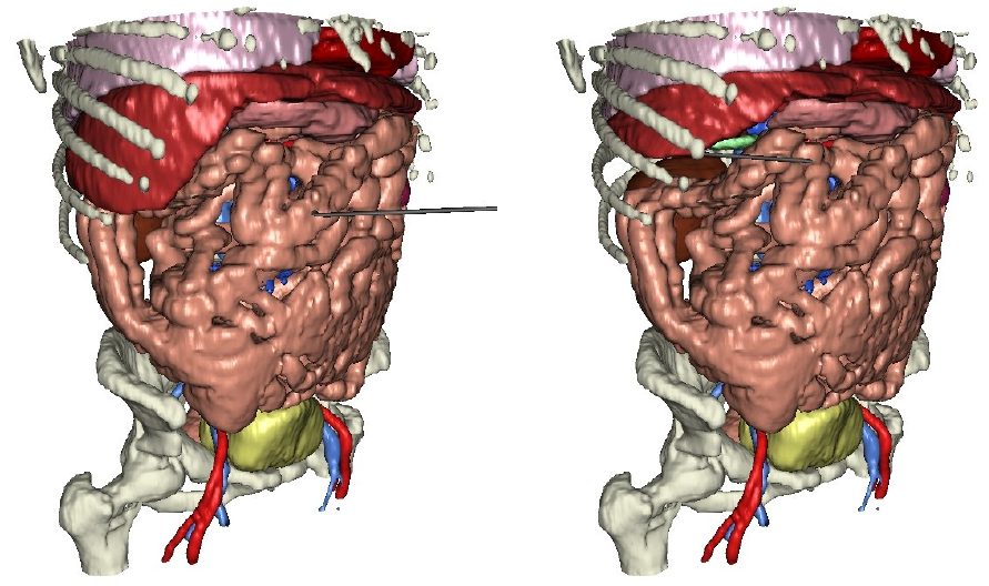 |
| Figure: Simulation of a patient-specific model built using medical images. |
Goal
The
goal of this work is to create a model of the peritony surrounding a
given liver and its surrounding organs. We will not consider the whole
peritony, but focus on the region of the hepatic pedicle. This video
of a real operation gives an idea of the geometry we target. Rendering
will not have to reach a high degree of realism in this work, since we
will focus on geometry. The geometry of the peritony in this area is
similar with a folded sheet with fat tissues inside the folds. The
geometric model will include, in addition to the big organs already
modeled (arteries, bile ducts), the folded sheet as a 2D surface and
the fat as 3D volume. The geometry will then be exported as a SOFA
scene. As far as possible, the anatomical knowledge will be expressed
in the MyCorporisFabrica anatomical database. Physical simulation will
probably be needed to model the complex folds of the membrane.
The validation will be done by simulating the cutting of the peritony in SOFA.
Work
The practical work may be organized as follows:
- Bibliography
- Understand the geometry of the target region
- Setup a geometrical model (the main part of the work)
- Export the model as a SOFA scene
- Apply a cutting simulation using the available SOFA methods
SOFA, MyCorporisFabrica, C++ will be used.
Organization
- From the beginning of February to the end of June
- Tutored by François Faure and Olivier Palombi
- Salary: about 380 €/month